-
Title
-
Eng
Mouthline Pigmentation Loss and Fisheries Associated Injuries of Rough-Toothed Dolphins (Steno Bradensis) in Hawaii
-
Date
-
2017
-
Creator
-
Eng
Welch, Jennifer
-
Subject
-
Eng
Environmental Studies
-
extracted text
-
MOUTHLINE PIGMENTATION LOSS AND FISHERIES ASSOCIATED INJURIES OF
ROUGH-TOOTHED DOLPHINS (STENO BREDANENSIS) IN HAWAII
by
Jennifer Welch
A Thesis
Submitted in partial fulfillment
Of the requirements for the degree
Master of Environmental Studies
The Evergreen State College
December 2017
�© 2017 by Jennifer Welch. All rights reserved.
i
�This Thesis for the Master of Environmental Studies Degree
by
Jennifer Welch
has been approved for
The Evergreen State College
by
______________________
John Withey, Ph.D.
Member of the Faculty
______________________
Date
ii
�ABSTRACT
Mouthline Pigmentation Loss and Fisheries Associated Injuries of Rough-Toothed Dolphins
(Steno bredanensis) in Hawaii
Jennifer Welch
Long-term photo data provides details on movements, reproduction, and environmental
impacts for monitoring populations of free-ranging odontocetes using a combination of visual
characteristics. Pigmentation variation within species assists researchers in individual
identification and age class estimations with evidence for distinction. Rough-toothed dolphins
(Steno bredanensis) of Hawaii exhibit apparent age-associated trends of pigmentation loss
around the mouthline, holding evidence of environmental and/or genetic influences necessary for
population conservation. Fisheries interactions in this region pose a threat based on depredation
events by false (Pseudorca crassidens) and pygmy killer whales (Feresa attenuata) and photo
evidence of similar interactions by rough-toothed dolphins. Relationships between age,
mouthline pigmentation loss (MPL) and apparent mouthline injuries (MLI) were assessed for
samples within 15 years of photo data of rough-toothed dolphins in Hawaii. Barnacles acted as a
proxy for evidence of interaction with fisheries operations and quantified per age class using
similar methods for false killer whales and pygmy killer whales in Hawaii. MPL was scored
based on the degree of pigmentation surrounding the mouthline, from 1 (no loss) to 6 (mostly
white). Adults had the highest level of MPL followed by sub-adults. The comparatively small
sub-adult sample had high variation in MPL scores. Fifty-one adult, sub-adult and unknown age
individuals showed MLIs via attached barnacles. No MPL or MLIs were evident for juveniles,
calves or neonates, which comprised less than half the sample of photographs with mouthlines.
The first study assessing pigmentation loss in rough-toothed dolphins is evidence that MPL
should be continued for age assessment alongside existing methods and current validation
techniques. The prevalence of mouthline injuries within the photos supports observations of
interactions with Hawaiian fisheries. Building upon this evidence by increasing the sample size
and injury/scar identification and record should inform current policy surrounding the protection
and conservation of small odontocetes in Hawaii contiguous with commercial and recreational
fisheries.
iii
�Table of Contents
ACKNOWLEDGEMENTS……………………………………………………………………....v
INTRODUCTION……………………………………………………………………………...…1
CHAPTER ONE: Literature Review………..…………………………………………………….4
Localized Populations………………………………………………….………….4
Current Issues and Research Question…………………………………………….5
Section 1: Age Assessment………………………………………………………………..8
Trends in Research Methods…………………………….………………………...9
Absolute and Relative Methods………………………………………………….10
Photography and Photo Identification…………………………………………...12
Fin Notches and Pigmentation…………………………………………………...14
Photogrammetry……………………………………………………………….…16
Section 2: Species Knowledge of Steno bredanensis…………………………………....18
Field Methods for Age Determination…………………………………………...21
Mouthline Pigmentation Loss…………………………………………………....23
Section 3: Fishery Injury Assessment……………………………………………………26
CHAPTER TWO: MPL and MLI of S. bredanensis (rough-toothed dolphin) in Hawaii……….29
Methods…………………………………………………………………………………..29
Results……………………………………………………………………………………32
Discussion……………………………………………………………………………..…36
Concluding Remarks……………………………………………………………..50
Bibliography…………………………………………………………………………..…52
iv
�ACKNOWLEDGEMENTS
Above all, I have learned about the time and accuracy required for cetacean research.
Photographs are not numbers, do not follow guidelines, and cannot be boxed or systematically
defined. Nature reveals patterns to us: cues researchers must recognize to protect, manage and
conserve vital populations of species. With this broad knowledge, my hope is this research
enlightens you on rough-toothed dolphins – provoking thoughts, concerns and questions about
the conservation of odontocetes in Hawaii and worldwide.
This research was made possible by Cascadia Research Collective; especially Robin Baird, Sabre
Mahaffy and the rest of the Hawaii team who I thank for their time and guidance. I am grateful
for the perspectives and teachings from MES faculty, especially my reader, John Withey. I
found continual support and encouragement from my family, friends, and community during this
process – all fueled by the wisdom, patience and unconditional love of the Creator.
v
�INTRODUCTION
Survival and viability of a species is dependent on habitat availability and the ability to
reproduce in that environment (Lande 1988). Evidence of a positive relationship between
ecosystem functioning and measures of biodiversity often provide conservationists a valuable
tool for assessing community health and resilience. Coastal communities must consider these
measures for taking appropriate actions around primary and secondary resource provisions
(Worm et al. 2006). The high adaptability and cognitive functioning of marine mammals
challenges the fishing industry and biologists seeking to protect these populations. Humans
reinforce interactions via direct and indirect food provisions and gear changes, while
simultaneously punishing acts via negative stimuli. To inform decisions amid survival for both
human and cetacean communities, pursuit of knowledge at the species level is essential.
Species viability, or status in relation to extinction, is a product of population level
reproduction rates and is assessed in numerous ways depending on the species (Ruggiero et al.
1994). Common research methodologies rely on age data to predict population growth (Barlow
and Boveng 1991). For marine mammal species, steps for assessing population status may
involve determining the species’ age(s) at sexual maturity, learning how to identify individuals
within this age class, and surveying a population for the abundance of individuals within each
age class (Brown et al. 1994). With some life history knowledge of the species, this process
gives researchers insight into the reproductive status of a population.
Conservation organizations often seek to protect spaces and communities with the
greatest taxonomic and species diversity, such as with the establishment of Marine Protected
Areas (MPAs). Protection from human disturbance in these areas, over time, consistently shows
positive outcomes for target species and communities alike (Mellin et al. 2016, Bossley et al.
1
�2017). The preservation of genetic diversity within populations also acts as a buffer in the face of
environmental change (Hooper et al. 2016, Mellin et al. 2016), which affects many organisms
living in aquatic environments. Adaptations to natural and anthropogenic induced fluctuations in
temperature, salinity, water level, prey availability, light, and turbidity are critical to survival.
For example, the genetic variation among salmon (Oncorhynchus spp.) populations in the Pacific
Northwest dictates their return to streams with slight differences in temperature and flow regime
(Hodgson and Quinn 2002) and supports viability in the face of climate change through varying
physiological and behavioral adaptations among populations (Quinn et al. 2001).
In the Hawaiian Islands, macroscale abiotic factors, such as oceanic upwelling processes
support greater diversity and community resilience to increases in sea surface temperatures
(Lourenco et al. 2016). Unfortunately, rapid or unexpected environmental changes, commonly
resulting from anthropogenic activities, sometimes override the capacity for positive individual
responses and lead to species extinction. As a diverse community of marine mammals, including
pinnipeds, odontocetes and mysticetes survive off the islands, research biologists strive to collect
data on populations in this region with the knowledge that preserving individual species will
contribute to the overall ecosystem preservation and resilience. The following study focuses on
two factors specifically concerning the conservation of an odontocete population among the
Hawaiian Islands. The first addresses the age assessment of this population and species and the
second turns to the impact of human activity on species viability as historic local fishing activity
continues to thrive culturally and economically within the region.
The lengthy gestation periods of cetaceans and their tendency toward long-term bonds
results in annual variation in reproductive rates (Taylor et al. 1987). In other words, age at
reproduction is not always indicative of annual reproduction rates for cetaceans and is not an
2
�accurate metric of sexual maturity or age. However, compiling a more complete representation
of ages is a beneficial step toward viability assessment and determining trends within and
between populations of cetaceans (McFee et al. 2012). Since the most accurate age assessments
result from data collected from birth onwards (Thompson et al. 1999), longitudinal studies are
imperative to research on wild populations for which information on individual births, deaths,
and movements between stocks is recorded. Although many ecological principles hold for
cetacean societies, research methodology must conform to the unique and sometimes,
unpredictable, social behavior of smaller odontocetes.
Human well-being among the islands is also imperative, creating the opportunity for
partnerships between research biologists, policy makers and long-line fisheries. Collecting
evidence of interactions between marine mammals and angling activity helps to maintain the
fishery, the marine mammal community, the local economy and the overall ecosystem. This
study builds upon active research of these interactions through the investigation of injuries to
small odontocetes with adaptive forage strategies. The motivation behind this thesis is to
continue working with the available data, build upon and improve methodologies and enhance
species knowledge to inform decisions surrounding the conservation of rough-toothed dolphins
in Hawaii.
3
�CHAPTER 1:
Literature Review
Localized Populations
Behaviors are mediated by habitat preferences and provide a challenge for researchers
attempting to assess the age structure of populations. The aquatic productivity of the Hawaiian
Islands provides suitable habitat for rough-toothed dolphin (Steno bredanensis) and 17 other
documented species of odontocetes (Baird et al. 2006, 2008; Albertson 2016). Although less is
known about rough-toothed dolphins relative to other dolphin species worldwide (West et al.
2011), researchers are developing an understanding of the natural history of the population in
Hawaii. Site fidelity, or adherence to the islands, was a significant finding for this species,
overturning previous understanding of the solely pelagic distribution of rough-toothed dolphins
(Baird et al. 2008). Regional mixing from oceanic eddies and upwelling contributes to higher
productivity and residence of the population in Hawaii (Calil and Richards 2010, Lourenco et al.
2016). This fidelity allows researchers to establish a robust data set via more frequent encounters
with the population based on individual movement patterns and reward effort (i.e. distance and
time spent in the field for amount of data collected). Furthermore, the consistent presence of
cetacean populations in Hawaii is likely indicative of the abundance and diversity of lower
trophic level species (López et al. 2008). The designation of twenty Biologically Important
Areas1 in Hawaiian waters therefore not only assists stakeholders in the management and
protection of these populations, but protects the greater marine ecosystem of Hawaii (Baird et al.
2015).
1
Defined by region and species. For cetaceans, reproducibility and residency are included qualifying factors (Baird
et al. 2015).
4
�Current Issues and Research Question
Cetaceans inhabiting nearshore environments are susceptible to interactions with human
activities that may influence their behavior patterns. Hawaii is not only a biodiversity hotspot,
but an attraction for tourists, fisheries, research, and Navy sonar testing that may affect the health
and behavioral patterns of cetaceans (Blane and Jaakson 1994, Baird and Gorgone 2005, Baird et
al. 2008, Bradford and Lyman 2015, Tyne 2015). Few accounts of the effects of human activities
on the species exist in the literature due to fewer encounters with rough-toothed dolphins,
relative to other species. In Hawaii, however, tourism, fisheries, U.S. Naval activities and local
recreation all pose threatening opportunities for rough-toothed dolphins and other species (Baird
et al. 2008, Timmel et al. 2008). Evidence for these interactions includes associations with
recreational fisheries and fish aggregating devices in Hawaii (Baird et al. 2008), strandings and
incidental catches by lobster and finfish fisheries in Brazil (Monteiro-Neto et al. 2000),
interactions with Hawaii and American Samoa-based long-line fisheries (Nitta and Henderson
1993, NOAA 2014), and depredation in Angola (Weir and Nicolson 2014). The difficulty in
assessing behavioral responses and a small sample size resulted in less clear evidence of
responses of rough-toothed dolphins to Navy sonar in Hawaii (Baird et al. 2014). Though the
deep distribution of rough-toothed dolphins poses a challenge to research crews, Cascadia
Research Collective (CRC) prioritizes the collection of photo and behavioral data of this species
in Hawaii (Baird et al. 2006).
Photo data and behavioral observations are largely the result of opportunistic encounters,
which support research efforts contributing to the body of knowledge on rough-toothed dolphins.
Innovative and more frequent survey and identification methods assist data collection alongside
changes in movements or fidelity to regions of higher productivity (Ryan et al. 2014).
5
�Additionally, collecting data on highly mobile, free-ranging, marine organisms is a challenge.
Researchers often deploy tags or collect acoustical data on individual cetaceans to gather
information on migrations, diving behavior, habitat use, and responses to changes in their
environment (Díaz López and Shirai 2010, Baird et al. 2011, Baumann-Pickering et al. 2016).
Biopsy data is collected for studies focused on social and genetic structure (McSweeney et al.
2007, Baird et al. 2011, Jefferson et al. 2012, Silva et al. 2015). These methods cannot, however,
provide direct data on age. Therefore, when photographs from the birth year are unavailable,
researchers utilize other characteristics and behaviors exhibited by rough-toothed dolphins for
determining age. Characteristics include, sexual dimorphism (adult males larger than females),
fetal folds, scarring, relative size, and positioning (e.g. neonates and calves often swim close to
the mother in the ‘echelon’ position allowing researchers to distinguish these individuals from
juveniles) (Addink and Smeenk 2001). Furthermore, the unique physical phenomenon of
pigmentation loss around the mouthline, captured in photo data of some individuals in Hawaii
could provide information on age.
A preliminary review of photographs from 2003 to 2016 reveal potential age associated
pigmentation loss. The unknown cause of this pigmentation loss poses a broader question for the
population’s life history and interactions in Hawaiian waters. The environment provides
opportunities for scarring, such as inter-species interactions or opportunistic and accidental
encounters with fishing gear. Other environmental variables affecting immune response or
causing a biological response associated with skin pigmentation are also possible. Skin depigmentation characterized as vitiligo in humans, for example, may be an innate immune
response to cell stress (Ezzedine et al. 2015), potentially manifesting in other mammal species.
Pigmentation loss could also be a genetic (biological) response to age, such as the graying of hair
6
�observed in some terrestrial mammalian species. It is necessary to consider the interaction of
multiple causes which could be depicted by varied tones or patterns.
The potential for harmful and undesired interactions with Hawaiian fisheries, the need to
build on species information, the potential advancement in field methods and the available data
has collectively guided this research. This study 1) assessed potential trends in mouthline
pigmentation loss (MPL) with age and 2) quantified fisheries-related injuries per age class. The
photo data, CRC researchers and biologists, and the available literature suggested lower MPL
among the immature classes, from neonate to juvenile, than within mature sub-adults and adults.
The prevalence of injuries may be similar to recent results for pygmy and false killer whales in
Hawaii, but niche consideration, forage behaviors, prey type and distribution, will all influence
this outcome, described in the proceeding sections. Intra-species differences between males and
females for MLI and MPL is an additional variable to consider. Not much is known about
differences in foraging strategies or prey preferences between the sexes if they exist in Hawaii.
Male rough-toothed dolphin adults were found to feed on a lower trophic level than females in
French Polynesia (Kiszka et al. 2011). Scarring due to male-male aggressive interactions in
odontocetes is common for Risso’s dolphins (Hartman et al. 2016) and has been observed among
bottlenose dolphins (Scott et al. 2005). Although a freshwater odontocete, male Amazon river
dolphins (Inia geoffrensis), like rough-toothed dolphins show obvious sexual dimorphism and
pigmentation differences between the sexes. Martin and Da Silva (2006) observed more injuries,
scars (tooth-rakes), and pinker tones in males than females. Their sample was relatively large (n
= 378) given the status of this species suggesting sex-based genetic, behavioral, and
environmental components for these patterns.
7
�The extensive contribution of background that follows is meant to provide the reader with
a more comprehensive understanding of this thesis’ methods and outcomes. These research
topics expose valuable data available on the present population of rough-toothed dolphins in
Hawaii. Though individuals associate with specific islands and island regions, their environment
allows and suggests movement between areas and is not limited to Hawaii (Baird et al. 2008).
Therefore, though breeding may be seasonal and residency is confirmed for some individuals,
the rough-toothed dolphins in this study are spatially and genetically mixed with fidelity to the
Hawaiian Islands (Albertson et al. 2016, R. Baird, pers. comm.)
SECTION 1 – Age Assessment
The following section describes typical methods used for aging cetaceans, highlighting
significant benefits and limitations researchers have found with a focus on smaller odontocetes.
Age determination for cetaceans requires an exploration of the most accurate and precise
indicators, presenting challenges across species and research labs. The behavioral ecology of S.
bredanensis is one reason for the limited breadth of information on this species relative to other
delphinids, such as common (Delphinus spp.) and bottlenose dolphins (Tursiops truncatus).
Also, with cetacean research spanning non-profit, state, and federal organizations, and
competition for funding and publication, data on certain species, such as S. bredanensis may not
yet be available to the broader scientific and academic communities. Though not always a focal
research species, encounters with free-ranging groups of rough-toothed dolphins are increasingly
documented and reported near islands and coasts (Kuczaj and Yeater 2007, Weir and Nicolson
2014, Anoop et al. 2015, de Boer et al. 2016).
8
�Trends in Research Methods
Terminology within the literature on aging can raise questions if not clarified. Though used
interchangeably in the literature, age validation most clearly describes the process of confirming
age by referring it to a predetermined reliable source, established by validation from a previous
technique. For odontocetes, the most reliable sources, apart from long-term photo data, include
Growth Layer Groups (GLGs)2 in teeth and age at length measurements (Hohn, Lockyer and
Acquarone 2016). Age estimation encompasses the use of a method to present a numeric
indication of an individual’s current life span. These methods include photogrammetry and less
precise visual observations such as relative size, relationship (mother-calf), and pigmentation.
Species management requires data on correct age estimates of individuals within the population
(Barlow and Boveng 1991, Campana 2001): a challenging process for species with cryptic
behavior, threatened or endangered species status, high longevity, or those with environmental
barriers to human observation or sampling. Therefore, utilizing a variety of methods and
combining estimates generates the most reliable age assessments for cetaceans (Hohn, Lockyer
and Acquarone 2016).
Researchers have investigated and applied both relative and absolute aging techniques to
cetacean species. Relative age assessments include documenting associations with other
individuals (free-ranging), analyzing fatty acids, and aspartic acid racemization (Hohn, Lockyer
and Acquarone 2016). These methods are beneficial in cases where absolute age is unattainable,
but often require an accurate reference. This reference may be in the form of an absolute age
indicator, such as GLGs (odontocetes) or species-specific age at length measurements (Hohn,
2
GLGs are annual groups of one or two layers that appear in the cross sections of the teeth of some species of
odontocetes and other mammals (Sargeant 1959, Hohn, Lockyer and Acquarone 2016).
9
�Lockyer and Acquarone, 2016); data provided by captivity and incidences of by-catch (Hohn
1989, Siciliano et al. 2007). Once this information is accessible, researchers and biologists can
make age estimates by referring to the body of age literature. Literature containing variation in
locations and methods may strengthen or weaken true estimates. For example, determining
lengths at different ages using GLGs and direct measurements from stranding events at various
locations can provide an accurate age-length reference for future studies using photogrammetry
(p. 19). However, using only age-length data from the Caribbean to predict the ages of a
population in the northern Pacific might risk accurate estimations due to differences in
environmental variables that may influence growth. If reinforcing, a combination of
methodology, population differences, and individual specimen data provides the most robust age
estimates for a species.
Absolute and Relative Methods
Aspartic acid racemization (AAR) is a relative aging method that has had some success
for older species of cetaceans (Rosa et al. 2012, Hohn, Lockyer and Acquarone 2016). The
process of AAR is tedious, requiring careful disassembling of the eye lens, stable temperatures,
and contamination avoidance. Additionally, an absolute age via GLGs or photo identification is
necessary to calibrate and validate the AAR results. Although this technique can be used for both
mysticetes and odontocetes, rates are different between species (Hohn, Lockyer and Acquarone
2016). Fatty acid signatures (FA ratios) have also been successful for finding the relative age of
toothed and baleen cetaceans in the absence of long term data. This process requires a blubber
sample that can be easily obtained by most research teams using biopsy methods. The limitations
of this method for aging may include lack of comparative ability due to differences in diet and
life histories. However, combining FA ratios for killer whales in the eastern North Pacific,
10
�produced a model able to estimate the ages of individual killer whales (Orcinus orca) within
±3.8 years (Herman et al. 2008). There may be risk of infection using biopsy darts, preventing
some researchers from using this method to age certain populations (Hanson 2016). CRC and
numerous other research organizations, however, have found this technique to be generally safe
for cetacean populations (Baird et al. 2013, Kowarski et al. 2014, Reisinger et al. 2014). Biopsy
sampling is currently used on rough-toothed dolphins in Hawaii for determining sex and genetic
variables, but not solely for aging. Relative to other aging techniques, researchers have classified
both AAR and FA ratios as costly and time consuming (Hohn, Lockyer and Acquarone 2016).
Scheffer published findings of layering within the cross section of bottlenose dolphins
(Tursiops truncates) teeth in 1950, similar to otolith and scale ring formation in bony fish
(Campana 2001) or (xylem and phloem) within tree trunks (Arno and Sneck 1977). Numerous
bottlenose dolphin studies, thereafter, confirmed the annual deposition of dentine layers: true to
actual age and collectively named “growth layer groups (GLG)” for the variation in within-year
layering patterns (Sergeant et al. 1959). Past uncertainties of this method included inter-species
calibrations (i.e. Is the method applicable across species of dolphins?) and variation in results
across studies due to differences in the process of determining the number of layers (Hohn,
Lockyer and Acuarone 1989). However, a number of studies confirmed similarities in GLGs
across species of odontocetes allowing for its broader application (Hohn, Lockyer and
Acquarone 2016). Although procedural differences across studies remain a concern that should
be considered before accepting the results as a reliable aging method for a species or across
species (Hohn 1989), using GLGs to find the absolute age of many odontocete species and
validate other aging methods is a most reliable and consistent method in current cetacean
11
�research. The field method of photogrammetry3, for example, is validated by comparing field
measurements to prior age-length data based on GLGs (Chong and Schneider 2001, Webster et
al. 2010). Determining age with lengths for rough-toothed dolphins was validated using GLGs in
Japan, Hawaii, and Brazil (Miyazaki 1980, West 2002, Siliciano et al. 2007). Researchers must
determine ages of multiple free-ranging individuals to get a clearer picture of the age distribution
of the whole population.
Photography and Photo Identification
Based on 2016 to 2017 data, CRC has identified over 2,300 rough-toothed dolphin
individuals among island areas of Kaua‘i-Ni‘ihau, O‘ahu and Hawai‘i. Long term photographic
records can describe an individual's’ age without the influence of environmental parameters such
as health or diet if taken within the first year of life. The general ease of photography as an aging
method and phenotypic characteristics captured in the photos can assist other methods of aging.
Since the beginnings of field studies on free-ranging cetaceans, photography and videography
have grown integral to research for a variety of applications (Bigg 1982, Hammond 1990,
Thompson and Hammond, 1992, Dahlheim, 1994, Mocklin et al. 2012, Weller et al. 2016).
Photography is used for population assessments and habitat use via photo identification data that
can validate residency of individuals and communities (Mayr and Ritter 2005, Parsons et al.
2009, Weller et al. 2016). This information advises local management and policy on, for
example, issues associated with fisheries (Forney et al. 2011, Baird et al. 2015). Therefore,
obtaining photos of high quality is a specific goal of this method and often distinguished by a
rating system within cetacean research (Baird et al. 2008, Kiszka et al. 2008, Urian et al. 2015).
In the field, researchers attempt these standards with permits to move within a certain proximity
3
Refer to section 1, page 19 for explanation
12
�to the animals, followed by maximizing the number of photos and photographers during an
encounter. These strategies increase the chance that at least one photo can be used for
identification and/or mark-recapture.
The regular surfacing behavior of dolphins allows researchers to visualize the unique
notching patterns and overall shape of dorsal fins helping to establish this structure as the
primary target for photo-identification. Digital sorting and rating of photos eases data review and
selection for multiple simultaneous projects when research species are many and effort distance
and time are large (Kaschner 2012, Baird 2013). In the first published photo-identification study
of rough-toothed dolphins, researchers focused on the stability of certain characteristics, finding
pigmentation and dorsal fin shape to be the most reliable (Mayr and Ritter 2005). Photographs
targeting specific regions of the body for identification may capture other physical characteristics
such as injuries, lesions, scarring, or parasitic organisms. These images reveal details on the life
history of an individual or population. Some rough-toothed dolphins in Hawaii show scars from
cookie-cutter shark bites providing evidence for their deep-diving behavior. False killer whales
often engage in depredation of long-line fisheries in Hawaii which is evidenced in photographs
of higher relative “dorsal fin disfigurements” (2000-2004) and mouthline injuries (Baird and
Gorgone 2005, Beach 2015), giving insight into their diet and ability to learn new foraging
techniques. Similar injuries have been noted for rough-toothed dolphins, but photo evidence is
still inconclusive on the prevalence of these interactions (De Boer 2010).
The maintenance and improvement of photographic techniques is critical for capturing
these unique features. Long term studies of cetaceans allow a history of photo data on individuals
to accumulate over time, providing clear evidence for ages (Hohn, Lockyer and Acquarone
2016). Research organizations, such as CRC, often utilize opportunistic or alternative methods to
13
�collect photos, such as whale-watching tour boats and photographers not directly associated with
research (Mayr and Ritter 2005) When comprehensive photo data is not available for individuals,
photographs may still provide characteristics from which to estimate age.
Fin notches & Pigmentation
Fin notches are used widely in identification and may provide information on age. Fin
notches may be obtained in a variety of ways depending on the species’ behavior and
environment. Narwhals (Monodon monoceros) in the Artic do not have dorsal fins, but were
observed to acquire “nicks and notches” on the dorsal ridge portion of the body over their
lifetimes (Auger-Methe et al. 2010). The researchers postulated that some originated from
anthropogenic activities due to the nick or notch location near bullet wound scars. Furthermore,
nicks, notches, age, and pigmentation loss seemed to increase collectively in this study,
indicating potential visual cues for age estimation. In the first published photo-identification
study of rough-toothed dolphins, researchers focused on the stability of certain characteristics,
finding pigmentation and dorsal fin shape to be the most reliable (Mayr and Ritter 2005).
Baird and CRC researchers rely on fin shape, which includes notches for identification
and mark-recapture data for odontocetes in Hawaiian waters. During a study assessing the
frequency of returns to areas within the Hawaiian Islands by rough-toothed dolphins, Baird and
colleagues (2008) determined a rate for accumulation of fin notches by dividing the collective
number of years over which a subsample of individuals was re-sighted by the total number of
notch changes (gains, losses or modifications). From these calculations they determined a
notching rate of approximately one every 2.4 years. This rate of change could be used towards
aging techniques for rough-toothed dolphins. However, using a quantitative assessment of
notches alone to assess the age of individuals could be problematic due to “loss” of notches or
14
�the inability to recognize new notches because of their location. A qualitative assessment of the
photographs may be more reliable for determining age. It would be difficult to predict age from
dorsal fin change over time if no prior photos for an individual existed. This form of age
estimation would probably be of little value given no alternate or prior information about an
individual or population.
Researchers have more recently investigated scarring and pigmentation loss as an
indicator of age in odontocetes. Researchers collecting data on the Indo-Pacific humpback
dolphin in China, via carcasses and photographed strandings, found variation in pigmentation
patterns for six different age classes (Jefferson et al. 2012). They observed an increase in
spotting with age that began to diverge based on sex during the sub-adult stage. Using tooth
GLGs and size to justify the age of each specimen, the researchers determined that adults may be
spotted or unspotted. Here, the advantage of having individual specimens allows age to be
determined quite accurately (validated) by GLGs; equivalent to a record of photographs from
birth onwards for free-ranging dolphins. Denise Herzing uses spotting to determine the age class
of Atlantic spotted dolphins (Stenella frontalis) closely studied since 1985 in the Bahamas. In her
studies, the use of underwater cameras allows visualization of the entire individual, easing age
assessments of free-ranging dolphins via pigmentation (Herzing 1996, 1997). Similarly, recent
results of increased pigmentation loss due to scar accumulation could advance aging techniques
of free-ranging Risso’s dolphins (Grampus griseus). Although body scarring was evident for
both sexes, Hartman and colleagues attributed scar accumulation mainly to social encounters
involving teeth raking among males, increasing with age (Hartman et al. 2015). The “whiteness”
of these individuals was thought to act as a visual cue for male dominance. These studies
introduce variables which complicate, but specify the classification of free-ranging individuals
15
�based on both sex and age by looking at pigmentation. With data available on rough-toothed
dolphins, the second best single age predictor may be in the form of photogrammetry data.
Photogrammetry
Researchers use photogrammetry to measure organisms that are not possible to collect or
assess for direct measurements (Chong and Schneider 2001, Sironi et al. 2005, Leurs et al. 2015).
Simple photogrammetry involves taking photographs of two laser points, positioned a known
distance apart, that are projected onto the body of an animal (Durban and Parsons 2006).
Researchers also use stereophotogrammetry, which produces a three-dimensional image from
two cameras positioned a certain distance apart with a specific lens magnification (Brager and
Chong 1999). Photogrammetry is not a new development in cetacean research methodology, but
researchers are advancing apparatuses and exploring its use for different species (Cubbage and
Calambokidis 1987, Webster et al. 2010). These data collected directly or via laser
measurements, allow researchers to predict ages if previous lengths at known ages were collected
for the species. Eaton and Link (2011) used this method to predict the ages of dwarf crocodiles
based on head lengths that isometrically relate to body lengths. If length-at-age data exist from
captive, stranded, dead, or entangled individuals of the species, photogrammetry data from freeranging individuals can help predict their age class. Autopsy data, for example, allowed Webster,
Dawson and Slooten (2009) to use photogrammetry to find fin width to be a reliable predictor of
length, allowing them to determine the age class of free ranging Hector’s dolphins
(Cephalorhynchus hectori) in New Zealand waters.
CRC has used photogrammetry on various species; in Hawaii, most notably on false killer
whales. Photogrammetry data for rough-toothed dolphins is currently limited, but more
opportunities to use this technique on this species in Hawaii could build a database useful for age
16
�assessments. CRC does not have readily available data on direct measurements and age of these
individuals, but the literature provides some information that may be used as a reference for field
length measurements. Siciliano and et al. (2007) aged 20 rough-toothed dolphins from museums,
strandings, and victims of fishing activities off south-eastern Brazil using lengths and GLGs.
Using the Gompertz model, they determined growth of S. bredanensis to stabilize at 258.1 cm
and 10 years. The majority of individuals were male and all were considered adults (> 15 years).
The stranding events of three individuals off Washington and Oregon of the United States
allowed researchers to determine the minimum ages (GLGs) and lengths of two males and one
female: 209 cm and 14 years, 192 cm and seven years, 219 cm and five years, respectively
(Ferrero, Hodder and Cesarone 1994). Results from studies on rough-toothed dolphins from
Japan measured males of 14 years at 225 cm and females of 10 years at 210 to 220 cm in length
(Perrin and Wursig 2009).
Photogrammetry has some limitations. Leurs et al. (2015) found body curvature to be a
potential issue after attempts to improve accuracy by changing the distance between the two laser
points on the animal. Prior to use, they tested their method for differences between
photographers, angle of reference, and inaccurate lens readings. Potential advancements for field
measurements require thorough calibration. Some studies have utilized a physical model of the
animal, taking photographs at multiple angles and distances to ensure best accuracy (Chong and
Schnider 2001, Webster et al. 2010). Environmental variables on the water, such as waves and
glare are sometimes unavoidable, therefore making the assurance of quality photographs for
analysis key to reliable data. Webster and colleagues (2010) noted the inevitability of errors in
photogrammetry since free-ranging dolphins are generally in constant motion. Furthermore, even
stranded or lifeless carcasses could introduce bias due to physiological differences or
17
�gravitational pull from terrestrial position. However, lengths at different life stages have some
degree of variation based on individual differences. For these reasons, it is necessary to take
precautions when utilizing new technology for data collection. By combining two or more of
these aging methods, researchers can ensure greater confidence in their results and more reliable
evidence for conservation.
SECTION II - Steno bredanensis Species Description
The general knowledge of rough-toothed dolphins as a species is still in the beginning
stages compared to other cetaceans. Information on life history, genetics, population structure,
habitat, and behavior are generally specific to certain regions and populations, contributing to
their “researchability”. As cetaceans learn new foraging techniques based on differences in
human fishing technologies, rough-toothed dolphin mouthline injury appearance and prevalence
may be/become regionally unique. Furthermore, if MPL has a genetic or environmental
component, this phenomenon could appear different on a global scale. However, the genetic
similarity of individuals and populations within any species is apparent in significant overlap of
life histories that can be focused on to increase species knowledge. When differences are
considered and addressed, small sample sizes may be combined to help develop a more robust
general knowledge of S. bredanensis and assist in its management worldwide (West 2002).
Reported group sizes of rough-toothed dolphins are variable and sometimes dependent
on dispersal, with widespread groups showing congregations of subgroups (Baird et al. 2008,
Ritter 2002, West 2002, West, Mead, and White 2011). West (2002) indicated smaller groups off
French Polynesia, where rough-toothed dolphins are encountered quite frequently compared to
the Eastern Tropical Pacific. Similar group sizes with wide ranges and frequency of encounters
18
�seem to occur off the Canary and Hawaiian Islands (averaging 16.8 and 7.0 individuals,
respectively) suggesting similar life histories between these populations (Ritter 2002, Baird et al.
2008). Barlow (2006) estimated a mean group size of 14.8 in Hawaiian waters spanning a
broader range. The productivity around these islands most likely lends to the attraction and
fidelity of these populations to them.
More frequent encounters near land masses have invalidated previous assumptions of
strictly pelagic, offshore habitat. In addition to Hawaii, individuals re-sighted around Utila,
Hondurous in the Caribbean may also be part of a resident population (Kuczaj and Yeater 2007).
Strandings occurring where sightings are uncommon support their offshore distribution (Ferrero,
Hodder and Cesarone 1994). Researchers have observed most individuals in tropical and subtropical regions including, but not limited to, West Africa (Addink and Smeenk 2001, de Boer
2010), Brazil (Siciliano et al. 2007), Hawaii (Baird et al. 2008), the Caribbean (Kuczaj and
Yeater 2007, West, 2011), the Mediterranean (Ryan et al. 2014), the Eastern United States and
the Gulf of Mexico (Waring et al. 2014, Wells et al. 2008). Although they are widely dispersed,
the National Marine Fisheries Service (NMFS) recognizes only three geographically based
“stocks” worldwide: Hawaii, Northern Gulf of Mexico, and Western North Atlantic (NOAA
2013, Waring et al. 2014). However, unknown or unreported genetic differentiation likely exists
within these stocks, as Albertson et al. (2016) found among the Hawaiian Islands.
Offshore and nearshore presence shows rough-toothed dolphins utilize shallow and deep
habitats with observations ranging from 20 m to 2,500 m depth (more generally between 100 m
and 1,000 meters) in the Canary Islands (Ritter 2002), 1,000 to 2,000 m in French Polynesia
(West 2002), and up to 4,000 m in Hawaii (Baird et al. 2013). Like other species, rough-toothed
dolphins have a flexible, fairly opportunistic diet, focusing on certain groups of prey. Recorded
19
�prey items include mahi-mahi (Coryphaena hippurus), houndfish (Tylosurus crocodilus), smelt
(Atherinops affinis), and squid (Ferrero, Hodder and Cesarone 1994, Pitman and Stinchcomb
2002, Baird et al. 2008b, West et al. 2011). Rough-toothed dolphins also pursue mackerel bait
and angler-targeted tuna (Thunnus spp.) in Hawaii (Nitta and Henderson 1993, D. Fleetham,
personal communication October 30, 2017). Long-term surface behavior allows researchers to
collect ample photo and biopsy data from S. bredanensis. Yet, the cohesive swimming patterns
of individuals in subgroups make capturing individual photographs a challenge. These surface
observations have enlightened researchers of the seemingly strong social structure and
associations between individuals; not unusual among delphinid species (Addink and Smeenk
2001, Mayr and Ritter 2005, Kuczaj II and Yeater 2007, Baird et al. 2008b). Observations of
rough-toothed dolphins with other cetacean species occurs and foraging may elicit associations
with various seabird species (Baird et al. 2008b). Individuals engage in bow-riding and display
curiosity by approaching ocean vessels and equipment (Kuczaj II and Yeater 2007), but show
more hesitancy than other delphinid species (Baird et al. 2008b, Jefferson 2009). Food reinforces
behaviors and outcomes associated with fisheries, such as depredation, entanglements,
interactions with aquaculture and trawling, and gear-related injuries; all potential threats to the
health and survival of individuals (Addink and Smeenk 2001, Baird et al. 2008, de Boer 2010).
The pigmentation of rough-toothed dolphins remains relatively stable over time and can
be used for identification purposes (Baird et al. 2008b). Individuals tend to be grey with slight
variations of brightness, scarring, and distinctiveness of the characteristic dorsal band between
age groups and individuals (de Boer 2010, Jefferson et al. 2006, Ritter 2002). An all-white
individual was sighted off Gabon, West Africa, but albinism could not be confirmed (de Boer
2010). Photos by Baird and colleagues from Hawaii indicate extensive belly scarring from
20
�cookie-cutter shark bites and various degrees of pigmentation loss on the ventral side and around
the mouthline. Observations of mouthline whitening outside of Hawaiian waters are limited or
under-reported. Addink and Smeenk (2001) made note of no whitish color “on the lips”,
suggesting individuals in other encounters off West Africa may have shown this characteristic.
During their encounter with a group of rough-toothed dolphins and melon-headed whales
(Peponocephala electra), Jefferson and colleagues (2006) noticed variable degrees of white
around the mouthlines of larger melon-headed whales compared to the smaller ones. Weir and
Nicolson (2014) published an image from an underwater video of rough-toothed dolphins during
a depredation event provided by crew members troll fishing off Angola. Some of the individuals
in the image seem to have white around the mouthlines indicating potential environmental or
genetic similarities between Pacific and Atlantic populations. Though more difficult to assess,
different pigmentation tones or patterns may be linked to region or temperatures; Jefferson
(2009) notes pinkish hues to areas of pigmentation loss in tropical locations. The more
observations such as these that are gathered, the better researchers can predict the prevalence
among populations, determining if it is a species-level attribute.
Field Methods for Age Determination
Age determination for wild rough-toothed dolphins may vary somewhat with researchers,
location, and organization. The common, reliable and consistent age determination method
worldwide is the knowledge of birth year using photo identification methods; this data can
validate any other methods developed for the aging of free-ranging individuals (Hohn et al.
2016). Since population estimates of rough-toothed dolphins are mostly unconfirmed where the
species exists, researchers encountering new individuals need a way to assess age. CRC staff
21
�currently use or have used: time in catalog (TIC), relative size (RS)4, association with other
individuals, and pigmentation to help determine which age class (neonate, calf, juvenile, subadult, adult) an individual belongs to. Fatty acid racemization (FA) is used if tissue samples are
obtained and requires lab work. Most researchers utilize similar subjective ‘sizing’ methods to
determine age class in the field (Addink and Smeenk 2001, Lodi 1992, Pitman and Stinchcomb
2002). Ritter (2002) identified individuals as juveniles that were “two-thirds” the body length of
adults. Individuals were recorded as calves if they were smaller yet. Kuczaj and Yeater (2007)
and de Boer (2010) identified the age class of the rough-toothed dolphins observed in their
studies, but do not describe their method of determination. Addink and Smeenk (2001) also used
the interactions and surface behaviors of mothers, calves, and “small juveniles” to identify their
relative ages. West (2002) defined a calf as “an animal that appears to be either newly born or
still maternally dependent” (p. 31). Not only do age estimations inform biologists and managers
of the reproductive status of dolphin populations, but are integrated into studies which determine
human impacts on resident populations (Krahn et al. 2009, Díaz López and Methion 2017).
The fidelity of rough-toothed dolphins to the Hawaiian Islands (Baird et al. 2008,
Albertson et al. 2016) makes it vital for researchers to continue to develop a body of knowledge
for the stocks in these waters. In 2006, there were an estimated 1,713 individuals in the Main
Hawaiian Islands and 6,977 in the Outer Exclusive Economic Zone (EEZ) (Barlow, 2006). CRC
staff have identified over 2,300 individuals in the waters surrounding Kauai, Ni’ihau, and
Hawai’i Island. The National Marine Fisheries Service (NMFS) of NOAA is responsible for
regulations pertaining to the protection and management of rough-toothed dolphins in Hawaii.
These decisions are based on NMFS research and that of contributing organizations such as CRC
4
RS can be used to justify an adult ten years later
22
�(NMFS 2004). The island fidelity these individuals display makes them a focal population to
also enhance the body of knowledge on S. bredanensis. The literature suggests age determination
is most accurate with a complete photo history for each individual: a challenging and expected
task to advance studies of identified individuals and document new encounters. It is therefore
appropriate that CRC utilizes a combination of technologies and methods, when feasible, during
longitudinal studies for proper management of cetacean populations. CRC maximizes aging
techniques using a combination of FA, RS, TIC, lips (ML pigmentation), pregnancy, calf
presence, and fetal folds.
The body of literature on pigmentation variation among different cetacean species and
between individuals, age groups, and sexes of the same species is relatively robust, but warrants
progress. No published research exists on the variation in mouthline pigmentation of roughtoothed dolphins. The availability of photo data from a consistent population of genetically
distinct rough-toothed dolphins in Hawaii provides a unique opportunity to investigate this
phenomenon and advance species and marine ecological research.
Mouthline Pigmentation Loss
A portion of individual rough-toothed dolphins photographed in Hawaiian waters display
pigmentation loss to varying degrees. Their gray to dark gray pigmentation allows this loss to be
visible as white or light pink random blotches or spotting, commonly observed on the ventral
side and around the mouthline. Other regions, such as the tip of the dorsal fin, depict
pigmentation loss. In general, these markings are produced by a few known, and potentially
unknown, mechanisms. Many cetacean species with darker pigmentation wear noticeable scars
due to the vulnerability of their skin to various interactions within their environment related to
socializing, foraging, and object encounters. In a physiological study, Lockyer and Morris
23
�(1990) found that bottlenose dolphins (Tursiops truncatus) re-pigment from minor wounds and
scratches within a year’s time. As expected, the deepness of the wound determined the scar
longevity: they observed semi-permanent scarring from teeth, otters, fishing gear and boat
collisions, gunshots, and shark bites. Rough-toothed dolphin skin similarly tends to re-pigment
within the lifetime, albeit within an unknown or unrecorded duration (R. Baird, pers. comm.).
These injuries give researchers an idea of the various interactions smaller odontocetes experience
in their lifetimes, informing wildlife managers and the public of necessary precautions and
regulations.
Social studies of multi-age dolphin “communities” also require researchers to know how
to indicate the ages of individuals. Without data available from birth, Lusseau and Newman
(2004) viewed scarring, size, and mother-calf associations underwater to assess age during a
study on the social structure of a bottlenose dolphin population in Doubtful Sound, New Zealand.
The authors claimed that cumulative scarring caused by aggressive interactions and sharks,
accumulated with age and was more apparent for male dolphins. Based on the previous study by
Lockyer and Morris (1990), the wounds that produced these scars would need to be deep or
recurring to slow or inhibit re-pigmentation, to be a reliable indication of age. Therefore, the
researchers’ decision to use a combination of age determination cues was thorough methodology
for this study. Both sexes of Risso’s dolphins exhibit scars from sharks, cephalopods, and other
same species individuals (Hartman et al. 2013) though significant differences were observed
between adult males and females due to inter-male aggressive interactions (Hartman et al. 2016).
Observations of the accumulation of scarring over time from teeth rakes between male Risso’s
dolphins have led researchers to hypothesize their contribution to a visual dominance hierarchy
(MacLeod 1998). Similar scar accumulation and scar tissue development from aggressive
24
�interactions between Amazon river dolphin males leads to noticeable discoloration, from dark to
pink, over time (Martin and Silva 2006). The social function of pigmentation loss could therefore
be used as an aging tool for researchers studying these populations (Hartman et al. 2015).
Foraging behavior and techniques may also contribute to scars and help explain
mouthline pigmentation loss (MPL) in rough-toothed dolphins. Various diving cetacean and fish
species obtain circular scars from cookie cutter sharks (Isistius spp.) that engage in diel
migrations (Moore et al. 2003, Papastamatiou et al. 2010, Sweeney et al. 2007). These scars on
individual Cuvier’s beaked whales (Ziphius cavirostris) and Blainville’s beaked whales
(Mesoplodon densirostris) in Hawaii are used for identification, age, and even sex (Sweeney et
al. 2007). Although sperm whales (Physeter macrocephalus) were once hypothesized to receive
scars on the head from squid, more recent research attributes these marks to aggressive male
social encounters (Whitehead 2003). In his book that corroborated studies on the life history of
sperm whales, Whitehead (2003) displayed a photograph of the white, lower jaw of an adult and
suggested its use in foraging to direct or distract prey at great depths. Werth (2004) examined the
tongue and jaw structure of sperm whale specimens, determining a suction mechanism and
potential luring purpose of the “white mouth”. Although, these studies give no description or
hypothesis for the lower jaw pigmentation pattern observed on adult individuals, its function may
have similarities to the whitish mouthlines of rough-toothed dolphins. Evidence does suggest that
North Atlantic humpback whales (Megaptera novaeangliae) lose pigmentation on the rostrum
over years of foraging for benthic prey (Canning et al. 2011). Repeated “scuffing” contributes to
the persistence of white scarring for a reported period of 12 years (Clapham et al. 1995). Canning
and colleagues (2011) found that this scarring was more apparent on older individuals within
their small sample, finding a significant difference between three age classes. Rough-toothed
25
�dolphins may show similar trends from foraging on large fish and/or contact with abrasive
objects associated with their prey.
Jefferson and colleagues (2006) observed melon-headed whales (Peponocephala electra)
and rough-toothed dolphins, for the first time, in a nearshore region around the Mariana Islands.
They noted white mouthlines, or “lips”, on some of the adult females and younger male melonheaded whales. The white around the mouths of individuals in the authors’ underwater
photographs of melon-headed whales does not seem to extend beyond a few centimeters around
the lips. These observations suggest that mouthline whitening could be environmental, rather
than, genetically based. However, the genetic based hypothesis for rough-toothed dolphin MPL
should not be dismissed since the extent and color variation (white to light pink) could indicate a
mix of genetic and environmental causes.
SECTION III - Fishery Injury Assessment
Evidence for marine mammal interactions with fishing operations exists from direct and
indirect observations which are often a result of learned and adaptive dolphin foraging response
behaviors to environmental change. Fishermen have reported common dolphins (Delphinus
spp.), bottlenose dolphins, rough-toothed dolphins and other odontocetes interacting with troll,
trawl, long-line, and purse seine fishery operations world-wide (Schlais 1984, Nitta and
Henderson 1993, Zollett and Read 2004, Forney et al. 2011, Ansmann et al. 2012). These types
of interactions are not limited to odontocetes: Humpback whales were observed routinely
foraging near hatcheries in Alaska upon the release of juvenile salmon (Chenoweth et al. 2017).
Recreational fishermen report sealions harvesting salmon directly from individual lines while
they retrieve their catches on the Columbia River, WA (personal communication with fishermen,
26
�September 2016 to 2017, Walker Jr. 2015). The economic loss for fishermen and the concern for
populations among biologists and conservation managers reinforce the reporting frequency
among both parties. In the United States, anglers and marine mammal biologists work together to
find solutions to issues such as depredation, but connections between stakeholders are weak in
some U.S. regions. This is concerning due to the high incidence of injuries and mortality due to
suffocation, entanglement and by-catch.
In Hawaii, fisheries interactions are apparent via direct observations by commercial and
recreational fishermen and photos capturing injuries, most commonly, on the dorsal fins and
mouthlines of a few species of small odontocetes (Beach 2015). These anatomical regions may
be more susceptible to contact with hooks and ropes from depredation, swimming near boats and
around nets. The attachment of barnacles to teeth after mouthline injury occurs assists
researchers with their identification. Notable incidences of barnacle growths occurring in
association with commercial fisheries include on a deceased striped dolphin (Stenella
coeruleoalba) in the Meditteranean, spotted porpoise in the eastern Pacific (Stenella graffmani)
and false and pygmy killer whales in Hawaii (Perrin 1969, Aznar et al.1994, Beach 2015). The
risk for disease and infection increases with deeper wounds such as these and could threaten the
reproductive health of the population if consistent for sexually mature individuals. White
scarring may not be an accurate predictor of the frequency or degree of fisheries interactions and
depredation for rough-toothed dolphins due to repigmentation. Additionally, pigmentation loss
around the mouthlines may mask scarring. Barnacle attachment, therefore, aids in quantifying
fisheries interactions, namely depredation, in rough-toothed dolphins.
The National Oceanic Atmospheric Administration reports injuries of rough-toothed
dolphins due to the shallow-set longline fishery that targets swordfish (Xiphias gladius). It is
27
�likely that mouthline injuries are a result of individuals targeting the required bait species,
mackerel, set at 30 to 90 meters deep. Established in 2010 under NOAA (2014), The False Killer
Whale Take Reduction Team reviews and assesses data, convenes with the public, and
establishes plans to reduce injury and mortality of odontocete interactions with fisheries in
Hawaii. Data and observations supporting the risk of interactions with false killer whale
populations have made this a prime species of concern. Since 2004, nine other species of
odontocetes, including rough-toothed dolphins, and humpback whales have all been injured or
killed as a result of the long-line fishery in Hawaii (Bradford and Forney 2016). Depending on
the prevalence and severity, these populations could experience threats to local survival and
growth and continual monitoring informs researchers of sustainable levels. Given past reports,
publications, and photo-evidence for potential harm to rough-toothed dolphins, the following
study will provide further evidence to build upon for remedial action to occur.
28
�CHAPTER 2:
Mouthline Pigmentation Loss and Mouthline Injuries of S. bredanensis
(Rough-Toothed Dolphin) in Hawaii
METHODS
Data Collection
A team of Cascadia Research Collective (CRC) biologists, researchers and associates
gathered all photo data utilized in this study during surveys conducted in marine waters of the
Main Hawaiian Islands (MHI) from 2003 to 2016 as part of multiple long-term studies of
odontocete populations in this region. Over these years, research vessels followed over 87,000
km of trackline during line-transect surveys5 for a systematic, yet broad scale effort to maximize
encounters with rough-toothed dolphins and other species (Baird et al. 2013; [October 2016
O‘ahu field project update]. Unpublished raw data). Water depths ranged from one to 5,000
meters. Rough-toothed dolphins were encountered most frequently off the island areas of
Hawai‘i and Kaua‘i-Ni‘ihau in water deeper than 1,500 meters, though sightings occurred above
and below this depth (Baird et al. 2008). Photographers aboard these vessels primarily targeted
dorsal fins for photo-identification purposes during surfacing behavior using film and digital
SLR cameras with 100-300-mm zoom capability (Baird et al. 2008). Additional CRC and
contributor photos used in this study captured multiple above-and below-water behaviors in
which the mouthline was visible.
CRC staff store and maintain the photo data used in this study on a computer database,
organized by species, location, encounter and individual identification number. The photos were
viewed and selected using ACDSee Pro 7, Microsoft Photos, and Windows Photo Viewer. The
5
As described by Baird et al. 2013 (pp. 254-255)
29
�individual ID, photo number(s), classified age, validation method, and sex, indicated on the mass
data sheet, were entered into an Excel file. The following analysis methods were then tested and
conducted on the photos in the database for this thesis and future work.
Mouthline Pigmentation Loss (MPL) Assessment
To determine age-related trends of MPL, ML photos for individuals were sorted into
folders by age resulting in the following sub-samples: adults (334 individuals; 1,472 photos),
sub-adults (117 individuals; 244 photos), juveniles (896 individuals), calves and neonates (29
individuals). Calves and neonates are separate age classes, but were combined due to photo
quantity and zero observed MPL during previous reviews. Only photos that captured 75-100% of
the ML of at least one side (left or right) were analyzed for MPL. Similar to methods used by
Hartman (2015) to assess pigmentation loss of Risso’s dolphins, MLs received a numeric score
from 1 to 6, representing the level of MPL: 1= no loss to limited loss, 2=limited loss to
noticeable loss, 3=noticeable loss to moderate loss, 4=moderate loss to mostly white, 5=mostly
white to complete white around lips, 6=complete white around lips to most of beak white. If both
sides of one individual in a single encounter had greater than 75% MV and were clear enough to
score, only one side was selected for analysis. All mouthlines were scored without knowledge of
age. Each file contained the original date and location of the encounter from 2003 to 2016.
Additional information, including sex, classified age, and island area for each individual from the
CRC S. bredanensis mass data sheet updated in October 2016, was also recorded.
Each encounter (photographic bout of an individual) was scored independently since
sightings of the same individual in consecutive years had potential for different MPL scores and
injuries. This further helped to note physical properties of a “full spectrum” of MPL per age
30
�class. This method holds true across all age classes if ML photos existed for individuals in these
age classes across years.
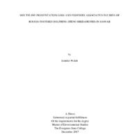
A
B
C
Figure 1. Examples of ML visibility percentage ratings. A. 100% of R side, B. 75% of L
side, C. 50% of L side. (ID and photo credits: A: HISb021. R. Baird, B: HISb0238. CRC, C:
HISb0276. Greg Shorr)
Mouthline Injury (MLI) Assessment
Mouthline injuries were assessed with methods like those used for pygmy and false killer
whales in Hawaii (Beach 2015) because of spatial overlap and potential interactions with the
same fishing equipment. The prevalence of MPL for many adults and the species’ repigmentation process made the visualization of vertical scarring, apparent in pygmy and false
killer whales, more difficult for rough-toothed dolphins in my personal experience. For this
reason, the majority of injuries were observed and noted by the growth of barnacles which is
indicative of a deep tissue wound (Beach 2015, Elorriaga-Verplancken 2015).
Photo files of over 2,300 rough-toothed dolphin individuals were viewed for mouthline
captures. The age classes adult, sub-adult, juvenile, calves and neonate were previously
determined by CRC using one or more of the following techniques or field age indicators: fatty
31
�acid analysis, relative size, time in catalog, lips (pigmentation), pregnancy, with calf, and
presence of fetal folds. Photographs which captured at least 50% of the total mouthline (left,
right and front), or had noticeable injury (attached barnacle), were placed in folders by
individual. Individuals for which multiple angles or sides were visible were considered for injury
analysis and assigned appropriate percentage visibility (e.g. 100% visibility of left side + 100%
visibility of right side = 200% total visibility). Beach (2015) found MV above 75% to positively
correlate with injury probability in false and pygmy killer whales6. Therefore, photos which
contained a high degree of interfering glare or water were eliminated and a higher selection
emphasis for 75% visibility and/or good quality reduced the number of photos to 3,309. The
percentage range of mouthline visibility (MV) for each age class was determined and compared
with total MLIs observed. Photo quality was visually assessed using prior established rating
protocol: 1=poor, 2=fair, 3=good, 4=excellent (Baird and Gorgone 2015, Beach 2015). Due to
the limited number of ML photographs within the data, percent visibility was assigned to the left
and right sides of each individual (Figure 1). Photos with lower than 50% visibility were used if
they had at least good (3) quality7. Quality rating was assigned by rating of the highest quality
photo if individuals had multiple mouthline photos for analysis.
6
7
This was done to increase probability of noting injuries prior to focusing solely on attached barnacles.
In results: Photos with a minimum of 10% MV were used.
32
�RESULTS
Over 73,000 photos and over 2,300 individuals were reviewed. Before a descriptive
analysis of the data, the sample for MPL assessment was filtered within the mass data sheet to
include individuals photographed ≥ 1 years; a step towards an improved, future method of
analysis described in later text. The resulting sample size for MPL analysis was 178 encounters.
A total of 51 individuals were found to have MLIs, though a greater prevalence is highly
probable within the sample based on unrecorded observations.
Mouthline Visibility
Mouthline visibility (MV) varied within each age class and across the entire sample. For
MPL analysis, all age classes except calves and neonates had a range of 75 to 100% MV (Table
1). A looser range to include individuals with 70% MV was applied to calves and neonates,
increasing sample size by only one individual. The initial review of all individuals within this
age class assisted the assumption that no MPL would occur here, lessening the concern of a
smaller sample size. MV for injury analysis of each individual included a combined calculation
of all available photographs during the single encounter for which the injury was recognized.
Since total MV for MLI assessment was not restricted to a 75% minimum, injuries were
recognized and recorded in encounters with a total of 10% MV.
Mouthline Pigmentation Loss (MPL)
Adults showed the highest range (5) and level of MPL indicating the greatest variation
within this age class, albeit with the largest sample (Table 1). A smaller sample of sub-adults
showed comparable variation to adults with a slightly lower range of 4, but had considerably less
MPL on average. Sub-adults had two maximum outlying MPL scores of five (Fig. 2). No MPL
was noticeable in the lower age classes.
33
�Figure 2. Boxplot of adult and sub-adult maximum (Q1), minimum (Q3), and
median MPL scores
Table 1. Mouthline visibility and pigmentation loss score results by age class.
Total
(n)a
Range of MV
(%)
Average MPL Scoreb
(mean, median,
mode)
Adults
137
75-100
4.5, 5, 6
5
Sub-adults
17
75-100
1.7, 1, 1
4
Juveniles
14
75-100
1
0
Calves and
Neonates
10
70-100
1
0
Age Class
a
b
Range of MPL
Score
Range of MV is presented for either the left or right side of each individual, not the total
MPL scoring: 1 – 6 (1: zero to minimal, 6: total loss around mouthline to mostly white beak)
34
�Important additional qualitative characteristics of MPL and irregular pigmentation
patterns included “piebald” individuals, as described by R. Baird (Baird et al. 2012), which had
similar appearing patterns of MPL. Similarly, in photos with the ventral side visible, individuals
with pigmentation loss showed similar, but more subtle patterns below the mouthline. These
areas appeared in subtle contrast with dark pigmented areas (Fig. 3A). When MPL was observed
it usually appeared most concentrated around the lips, with an upward reduction so the top of the
beak was still pigmented. Overall individual body pigmentation pattern and counter shading was
apparent in younger individuals, which made a significant portion of the mouthline appear
lighter, but different than the bright white contrast immediately around the mouthlines of many
adults. Some adults showed pigmentation loss solely on the lower jaw, below the mouthline.
This was apparent in individuals in which only the upper portion and none or part of the lower
jaw were visible in the photograph; not suitable for analysis, but important to note. Furthermore,
some MPL had a scar tissue appearance (Fig. 3D).
A
C
B
D
Figure 3. A) Ventral pigmentation loss that seems at an “advanced stage” future MPL, B) potential
teeth rake marks, C) areas of pigmentation appear raised, D) MPL appeared as scar tissue (Photo
Credits: A, B: Brenda Rone, C: Annie Douglas, D: R. Baird)
35
�Mouthline Injury Assessment (MIA)
The majority of observed injuries were indicated by attached barnacles (Table 2). As
previously mentioned, personal recognition of more minor injuries than barnacle sites resulting
from fisheries interaction were obscured by white mouthlines in adults. Therefore, only very
distinctive injuries were included. Attached barnacles were frequently in colonial formation, but
counted as a single injury unless observed on separate sides of the mouth/head.
Table 2. Number of individuals with injuries, quantity and type of injury by age class.
Number of Individuals
with Injuries
Range of MV (%)a
Quantity and Injury
Type (attached
barnacle – AB, fresh
abrasion – FA,
unknown – UNK)
Adults
48
10 - 195
46 ABb, 3 FA
Sub-adults
1
190 - 190
1 AB
Juveniles
2
90 - 200
1 AB, 1 UNK
Calves and Neonates
0
N/A
N/A
Unknown
1
100 - 100
1 AB
Age Class
a
MV range % sum of L and R sides
One individual had an AB on the L and R side, counting as two injuries.
b
Vertical line scarring, present on the lower, posterior mouthline (B) and behind the eye
(C) of individuals in Figure 2, similar to those observed by Beach (2015) on false killer whales in
Hawaii, were clear for over 10 individuals within the adult age class. These were most likely a
result of interactions with fisheries. In one instance, a rather large vertical mark was associated
with barnacles along the mouthline (Fig. 4c). Since calves and neonates nurse from their
mothers, the results for this age class are consistent with the highly unlikely instance of injuries
due to interactions with fisheries along their mouthlines.
36
�DISCUSSION
Mouthline Pigmentation Loss
The results were mostly consistent with predictions of higher MPL in the upper age classes.
A similar range, but lower average MPL among sub-adults suggests high variation among the
sub-adults. It seems MPL begins at some point within the sub-adult stage, but from these results,
is unclear how rapidly it progresses if it does, indeed, advance during this life stage. As
individual dolphins enter the sub-adult stage, they become independent from their mother and
generally form sub-adult groups that travel and potentially forage together (Wells 1991). The
inconsistencies of MPL, therefore, could be influenced by foraging style, associates, or
advancement within the group/age. Assessing a greater number of sub-adults for a more robust
average and a larger sample of juveniles would be a main focal point for improving this study
using the same methods. For now, field researchers may feel comfortable continuing to attribute
zero MPL to juveniles, calves, and neonate rough-toothed dolphins in Hawaii in combination
with other aging methods.
Pigmentation loss was more or less apparent based on the darkness of the individual’s
pigmented skin. Actual pigmentation loss may not be visible depending on this characteristic,
similar to the grayness of light colored or blonde hair with age in humans. The tone of areas of
MPL for some individuals appeared pink, rather than the bright white of most mouthlines with
MPL. It could not be determined if tone variation was due to natural light, the photograph,
biological or abiotic factors. Allen et al. (1993) found regional differences along the dark to light
spectrum of humpback whale (Megaptera novaeangliae) flukes among five northern breeding
regions and three southern breeding regions. The authors suggest differences in pigmentation a
visual indicator of genetic differences between northern populations. In some photographs of one
37
�individual during a single encounter, areas of MPL appeared pinkish or white, what appeared in
this instance to be a phenomenon caused by changes in natural light.
The possibility of some MPL to occur over time due to foraging is based on the premise that
the prey, or means of obtaining the prey, causes routine scarring. Rough-toothed dolphins
regularly feed on mahi-mahi, tuna, other bony fish and cephalopods in Hawaii (Baird et al.
2008b, West et al. 2011) Assuming consistent foraging behavior, any scarring may prevent
repigmentation around the mouth. However, with lack of evidence that prey items cause such
scarring, this hypothesis seems weak. It is important to mention, however, that Beach (2015)
noted both “natural” and foraging-related MPL patterns in pygmy and false killer whales in
Hawaii. Forage-related MPL in her sample was due to scarring from depredation and apparent by
white scars and “jagged, vertical cuts through the lip” in these species (Beach p.29). Unlike
rough-toothed dolphins, pygmy killer whales’ skin does not re-pigment making it easier to
distinguish and predict the cause of MPL.
Another potential cause of MPL is from the teeth of other individuals. Some of the images
showed teeth rake marks near or on the head region (Fig. 3B). Knowledge of rough-toothed
dolphin social behavior is minimal, based on publications. However, the aggressive intraspecific
interactions among male Risso’s dolphins, leading to intense scarification, may also exist within
rough-toothed dolphin social networks (Hartman et al. 2015). The CRC data provides the
opportunity for comparison between adult male and female MPL level and appearance.
Jefferson and colleagues (2006) observed melon-headed whales (Peponocephala electra)
and rough-toothed dolphins, for the first time, in a nearshore region around the Mariana Islands.
They noted white mouthlines, or “lips”, on some of the adult females and younger male melonheaded whales. The white around the mouths of individuals in the authors’ underwater
38
�photographs of melon-headed whales does not seem to extend beyond a few centimeters around
the lips. These observations suggest that mouthline whitening could be environmental, rather
than, genetically based. However, the genetic based hypothesis for rough-toothed dolphin
mouthline pigmentation loss should not be dismissed since the extent and color variation (white
to light pink) could indicate a mix of genetic and environmental causes. The data showed unique
patterns between adult individuals that may be assessed further. Just as human male adults
experience pattern baldness, a similar hypothesis may be proposed for rough-toothed dolphins.
Though studies on baldness in humans were inconclusive of genetic and hormonal associations,
Ellis, Stebbing, and Harrap (1998) determined genetics, age, and hormones to be broad
independent variables of differing patterns of hair loss that could assist postulations for roughtoothed dolphins. Other mammalian species, such as primates and mice, have also shown
patterns of balding, or alopecia (Novak and Meyer 2009). Odontocetes may be born with hair,
usually concentrated near the mouth, beak, and genitalia. Hair-like structures (vibrissae) with
thermoregulatory function remain for adult dolphins. Pigmentation loss associated with these
regions could be assessed with further knowledge of processes for rough-toothed dolphins in
addition to photo data: potential assessments addressing the relationship between sex, genetics
and pigmentation loss pattern. Future research to help understand general pigmentation loss
could utilize photos that display the ventral and lateral sides (Fig. 4).
39
�Figure 4. Ventral pigmentation loss of
adult HISb1075 (Photo credit: Brandon Southall)
A combination of genetic, behavioral and environmental variables is likely contributing
to MPL. Cell response to stress may be the cause of highly contrasting skin depigmentation in
humans, named vitiligo (Ezzedine et al. 2015). In humans, this disorder occurs across children
and adults, though some studies found increased prevalence with age. Oxidative stress, genetics,
and immune system responses are thought to act in concert for the loss of melanocytes in those
with vitiligo (Laddha et al. 2013). Further investigation of these causes for MPL in roughtoothed dolphins would most likely require tissue and environmental samples. It is difficult to
speculate causes of body pigmentation loss, but analyzing different regions (i.e. mouthline vs.
ventral side) could help researchers understand certain mechanisms contributing to these
patterns. Comparing Hawaii observations with international observations can help researchers
determine the breadth of MPL within the species S. bredanensis.
40
�Mouthline Injuries
Injuries resulting from interactions with fisheries were evidenced in this study by
colonized barnacles where mouthline tissue was broken. Although the MLIs recorded cannot be
completely attributed to depredation with absolute certainty, it is the best explanation of
causation. Similar to MPL, a greater sample size of sub-adults and juveniles would yield more
reliable results for the prevalence of MLIs within these age classes. Beach (2015) found one
barnacle attachment per 47 pygmy killer whales, or approximately 2% of the sample. Although
the observations in the present study for MLIs were encounters, not individuals, approximately
2% (48 ABs per 2,160 encounters) of the sample had barnacle attachments. Since the number of
encounters does not stray too far from the number of individuals viewed for MLIs (>2,000
individuals), these statistics increase speculation on the take of rough-toothed dolphins in
relation to the stock’s potential biological removal level. Depredation seems to be the sole cause
of most mouthline injuries, therefore noting the prevalence among the collective population of
rough-toothed dolphins in Hawaii is the primary and main concern for conservation and fishery
regulations.
The majority of individuals with MLIs and low mouthline visibility had the tip of the
nose above the surface. The location of these deeper injuries towards the front of the beak may
be indicative of the morphological differences (beak vs. melon) and depredation behavior or
strategy between rough-toothed dolphins and false killer whales. In only one instance, barnacles
occurred on the mouthline posterior to mid-way back from the nose (Fig. 5D). This observation
questions the prevalence of ABs occurring at or greater than 50% MV within the population.
Unsurprisingly, the frequency of MLIs among false and pygmy killer whales increased with MV
(Beach 2015), so this may be a detectability issue that a larger sample of rough-toothed dolphin
individuals with more than 75% MV could address. Additionally, this individual also had the
41
�highest density of barnacles along the right side making the number of injuries difficult to assess.
The majority of barnacle attachments can be attributed to depredation based on how roughtoothed dolphins hold fish in their mouths from above and underwater photographs (Fig. 5A, B)
and previously described observations of depredation. Recreational anglers off Angola witnessed
rough-toothed dolphins depredating from their fishing lines by “mouthing” and removing fresh
and plastic baits (Weir and Nicolson 2015). Though the gear and fishing method here are
different than long-line operations in Hawaii, photographs and videos from these types of
encounters are informative for future identification on MLIs. Furthermore, knowledge of
barnacle life-history would assist in determining the severity, timing and quantity of injuries
where barnacles are located along the mouthline. Since barnacles live in colonies, an “attached
barnacle” could include more than one barnacle on one injury, or one colony on multiple injuries
(Fig. 5C). One of 82 striped dolphins stranded in the Mediterranean had the crustaceans Lepas
pectinata, L. cf. hillii and Conchoderma virgatum attached to its teeth. Interestingly, the
researchers did not find any sign of damage that would indicate an injury (Aznar et al. 1994).
This observation questions the reliability of barnacles to act as a proxy for mouthline injuries in
future analyses. A more thorough qualitative approach to scarring and disfigurations among
rough-toothed dolphins before continuing to attribute all barnacle attachments with injuries
would be wise.
Although rough-toothed dolphins are more frequently sighted in deeper water than false
killer whales in Hawaii, spatial overlap occurs (Baird et al. 2008) and depredation from fishing
vessels navigating similar tracts is plausible. Marine mammal observers aboard fishing vessels
help quantify interactions and species. NOAA researchers noted two observations from the
Hawaiian Island shallow and deep-set longline fishery and six from the American Samoa deep-
42
�set longline fishery of rough-toothed dolphin interaction (death or injury) from data collected by
the Pacific Islands Region Longline Observer Data (PIRO) from 2009-2013 (Bradford and
Forney 2016). Yet, it is likely some observations of depredation go undocumented, especially in
areas without observer programs. In his book, The Lives of Hawai‛i’s Dolphins and Whales
(2016), Baird notes attempts of deterring rough-toothed dolphins via shooting or tossing hooked
fish at them. Researchers from CRC routinely observe rough-toothed dolphins swimming near
and around fish aggregating devices (FADs) such as large buoys (Fig. 6b). Since plankton and
bivalves accumulate on suitable surfaces, this food source attracts small fish which are preyed
upon by larger fish (Iglesias 1981). Similar to aquaculture operations (rafts, net-pens), dolphins
in Hawaii learned to locate and return to these stationary devices to forage (Diaz-Lopez and
Methion 2017). Perhaps injuries could be observed more closely in less stressful conditions at
locations such as these.
A
B
[
T
y
p
e
a
q
u
D
Co
t
e
f
r
o
m 5. Barnacle location seems consistent with prey acquisition and
Figure
mouthline
injuries for acts of depredation on long-line fishing gear.
t
(Photo credits: A: Deron Verbeck, B: Elisa Weiss, C: Dan McSweeney,
h
D: A. Douglas)
e
d
43
o
c
u
m
�a
b
c
d
Figure 6. Evidence of interactions between fisheries and rough-toothed dolphin in Hawaii:
a) Two individuals within proximity to a local fisherman, b) an individual near bouys that
attract fish (FADs), c) an individual swimming with derelict fishing gear attached and d) an
individual being sampled by CRC near floating derelict fishing gear. (Photo credits: a: Daniel
Webster, b: Julie Steelman, c,d: Jessica Aschettino)
Limitations and Future Research
It is important to consider the limitations of this research from data collection to analysis.
Cetacean field data collection is layered with challenges. The opportunity to photograph
individuals is dependent on dolphin behavior and weather conditions, limiting data collection to
certain days or times of the year. Since pigmentation seems to remain relatively constant within
years, data is mostly limited by behaviors for which the head is above the water. Though
behaviors are not discussed in depth here, individuals were engaging in surface and aerial
behaviors for the majority of these photos (raising head out of water while breathing, various
jumping, and feeding) while a few, clear enough for analysis, were captured underwater (Kuczaj
II and Yeater 2007, Ferrer-i-Cancho and Lusseau 2009). As Addink and Smeenk (2001) also
found, the close, synchronous surfacing of rough-toothed dolphins challenge photographers to
44
�capture individual photographs and some of these were found within the data. Though dolphins
may attempt to dislodge organisms via various styles of jumping, MLIs cannot be reliably
associated with one of these behaviors, avoiding any known bias. However, it is important to
note the potential for individual (personality) or age differences associated with raising the head
out of the water in any one of these behaviors. Fortunately, CRC associates are able to capture
underwater photographs that often provide clearer supplemental or primary data. Sunlight,
waves, underwater lighting, and angle to camera all distort the perception of MPL and
photographs in this study were influenced by these factors to some degree. In some instances
glare posed a problem for determining the level of MPL (Davis and Grayson 2007) in that more
glare or light focused on the mouthline highlighted gray areas creating the illusion of greater
pigmentation loss than was actually occurring (Fig. 6). This can be manipulated with ACDSee
Pro or other photo viewers, but could be problematic for immediate field assessment if methods
of MPL were to be used in this manner. Developing a larger sample size of high quality photos
should be addressed more acutely in following studies of MPL and MLI of rough-toothed
dolphins.
Figure 6. Left) Light can distort regions of MPL. (Photo credit: D. Verbeck), Right) A summarizing snapshot of the
clear distinction between age class using MPL as an indicator. (Photo credit: G. Shorr)
45
�Secondly, the initial and attempted method approach was determined based on the
prediction that the data would contain a low number of individuals photographed over multiple
years for which at least 75% of the mouthline was visible (preferably the same side) for each
consecutive year. However, determining averages for broad categories of age class had certain
challenges that could be avoided using the former method of selecting individuals photographed
preferably over the course of four or more years. For instance, it was necessary to avoid
individual differences by using each encounter as an observation. If observations were chosen of
the same individual in one year, one of those had to be removed from the sample so the
individual would not influence the results. Ultimately, this decreased the sample size and glossed
over variables that could be helpful considering rough-toothed dolphins as a wild species with
high variability and multiple innate and environmental influences.
Another limitation to sample size occurred from approaching the data (individuals in the
catalog) chronologically. This decision was made as a straight forward way to randomly choose
individuals, based only on the day photographed since individuals are given ID numbers as
sighted during fieldwork. The adult age class sample grew faster than the other age classes, so
conscious selection of lower age classes was necessary later during the review and sorting
process. Placing all individuals in categories of age class prior to reviewing for mouthline
injuries would be a more efficient way if this method is repeated in future research.
46
�Approaching the data from a mostly qualitative perspective was beneficial for this initial
analysis for multiple reasons. During some encounters, over 75% of both the left and right sides
of the mouthline were photographed. This increased qualitative data for how MPL may occur,
but this was an additional consideration during data compilation and analysis. In some instances,
both sides showed equal levels of loss (i.e. L 6, R 6), but others showed variation that equated to
close, but unequal scores for both sides (i.e. L 5, R 6). Since different scores meant differences in
MPL for one or multiple reasons, one of these sides was ignored when determining averages for
each age class; another instance of ignoring important data for this population of rough-toothed
dolphins and the species. However, more photos of both sides could shed light on these
inconsistencies and causes of MPL (Fig. 8).
Figure 8. Even with the majority of the mouthline visible above the water,
assuming uniformity of the entire mouthline would be a form of extrapolation
(ID: HISb0860). Individuals for which mouthlines were captured on both sides
and head provided evidence for non-symmetrical pigmentation loss between
the left and right sides – a factor that must be taken into consideration for
field and photo age predictions. (Photo credit: J. Aschettino)
For mouthline injury assessment, immediate future endeavors include expanding the
sample size and identification and categorization of mouthline injuries. It is likely that some
47
�injuries were overlooked; especially minor (less-severe) ones without barnacle attachments that
would heal faster. Also, as confidence increases for correlations between mouthline visibility
(MV) and pigmentation loss score, percent visibility could be lowered to include a greater
sample size for overall age determination. Determining how this approach would advance
research would need to be considered.
Cascadia Research Collective demonstrates thorough and opportunistic data collection,
maintaining date, source/encounter #, encounter code, cluster position, area, island,
original/within day or year/between-year sighting, span of years seen, # of years seen, # of times
seen, distinctive rating, # notches, notch location, scar/pigment marks, # photos, photo quality
ranking and side, tag or biopsy, tagged full sighting history, skin sample #, tag #, haplotype (y/n),
age/sex, temp ID #, and additional comments in addition to photos, for each individual per
encounter. Associations of MPL and MLI with island or other scarring and pigmentation marks
could provide further insight into their behavior and environmental impacts. If a greater number
of individuals are assessed for MPL, this data may be compared against fin notching,
photogrammetry measurements, and photographic history to enhance aging methods and better
understand the process(es) behind MPL.
Identification of injuries can be a challenge since not all depredation techniques are
known or have been witnessed in Hawaii. Furthermore, the healing process of rough-toothed
dolphin mouthline injuries is uncertain. Zasloff (2011) notes the unique and fast healing process
of bottlenose dolphins due possibly to the presence of antimicrobial isovaleric acid in the
blubber. It is an interesting case that such compounds are less prevalent in other marine
mammals (Kiipman et al. 2003). If other delphinids, such as rough-toothed dolphins, have
similar blubber composition to bottlenose dolphins, this would support the behavioral and
48
�physiological adaptability of this marine mammal family. With the continuing long-term
research of rough-toothed dolphins in Hawaii, potential research endeavors could include
targeting mouthline injuries over time to assess the healing process.
Additional and Alternate Method Approaches
A variety of other methods for examining MPL exist. Approaching the data with Bayesian
statistics and human rater participants, as Hartman et al. (2015) did for Risso’s dolphins could
add to reliability and consistency among rough-toothed dolphin researchers. Such analyses
would require a collection of adults photographed from birth onwards and/or a length at age
chart from CRC photogrammetry data and global evidence from the literature (some of which
was previously discussed). Certain limitations within the present study, including time and
uncertainty around the cause of MPL, may be addressed and a method approach similar to
Hartman et al. (2015) could be tested.
Researchers have also been experimenting with methods for the quantification of
pigmentation patterns on various taxa including, amphibians (Davis and Grayson 2007, Pokhrel
2009), insect larvae (Davis et al. 2004), raptors (Bloom and Clark 2001, Liguori 2004), and lions
(Miller et al. 2016). With the previous knowledge that phenotypic traits vary with age for
African lions, Miller and colleagues (2016) used photos to determine age classes that may be
recognized by hunters. Scores for the percentage of nose darkness in photos were compared
between students and an author and also quantified using Adobe Photoshop CS6
v.13.0.6darkness. They compared qualitative and quantitative values and found that the age of
free-ranging, large mammalian individuals could be distinguished by experienced hunters and
used for species management (Miller et al. 2016).
49
�The analysis of digital photos using different image analysis software is often useful
when there is sharp contrast between the color of interest and background pigmentation. Davis et
al. (2004) determined the amount of black pigmentation within bands on monarch caterpillars by
converting images to a black and white color before running them through a computer analysis
program. However, after the current analysis, attempts to quantify pigmentation loss may fall
short of reliable this early in research of MPL of rough-toothed dolphins: The variation between
individuals, tones and patterns suggest the working of multiple variables that may include
genetic, environmental, individual, sex, and age.
Statistical data provides numerical evidence for any trend in mouthline pigmentation
change with age: something that cannot be reliably determined by the human eye alone. A
quantitative procedure, such as determining pixel ratios, would seem to provide a more accurate
assessment of MPL with age, but could miss real variation. Furthermore, this slightly assumes
that skin color change around the mouth has a genetic or environmental cause that shows a
steady trend over time. For example, age has a certain degree of consistency, or pattern, over
time, for living things, which may be genetically linked to pigmentation changes, whether this is
a gradual or punctuated process. A qualitative analysis takes additional visual characteristics into
consideration, like color and solidness, which could have two different causes. For photo
identification, research maintains that the most accurate assessment of photographs is via
humans within the field (Gilman et al. 2016, Hartman et al. 2015). It is important to still consider
expert human judgement of age class recognition via pigmentation for field and lab work if
researchers and staff are provided photographs for comparison between age classes and given
training. Though things can be learned through this analysis that may assist future computerized
or visual aging methods, the core purpose of this study was not the formation of an aging model
50
�or method, but to determine if and how mouthline pigmentation relates to age. Like all studies,
these results are meant to be built upon by further study of the population of S. bredanensis in
Hawaii and elsewhere.
Concluding Remarks
The potential for more qualitative assessments for cetacean pigmentation patterns is
demonstrated with this research. The preliminary review of this data reveals a correlational, yet
complex phenomenon of pigmentation loss over time for rough-toothed dolphin in Hawaii. The
overall conclusions open avenues for multiple data assessment options. Individual differences
such as patterning of MPL around the mouthline, extending to the entire beak, may be interesting
given more photos including these regions. Future analyses noting the characteristics of MPL,
such as spread or density of “whiteness” may help researchers understand the primary causes
more clearly.
This research was accomplished with the effort of long-term data collection that must be
continued for the conservation of marine mammal species. Twenty years of data revealed greater
stability and response to disturbance events within designated MPAs of the Great Barrier Reef
than unprotected areas (Mellin et al. 2016). Additionally, twenty-four years of survey data
showed the abatement of various environmentally detrimental human activities within the Port
River estuary of Adelaide, Australia to correspond with more frequent nearshore bottlenose
dolphin (Tursiops aduncus) sightings (Bossley et al. 2017). This fate is especially true for
specialist and long-lived species who depend on certain habitat characteristics for survival and
viability. For rough-toothed dolphins and odontocetes collectively, the hope is that this research
may suggest research hypotheses, expand methods and inform local and international research
and conservation.
51
�BIBLIOGRAPHY
Allen, J. M., H. C.Rosenbaum, S. K. Katona, P. J. Clapham, and D. K. Mattila. 1994.
Regional and sexual differences in fluke pigmentation of humpback whales (Megaptera
novaeangliae) from the North Atlantic Ocean. Canadian Journal of Zoology 72:274-279.
Albertson, G. R., R. W. Baird, M. Oremus, M. M. Poole, K. K. Martien, and C. S. Baker.
2016. Staying close to home? Genetic differentiation of rough-toothed dolphins near
oceanic islands in the central Pacific Ocean. Conservation Genetics 1-19.
Ansmann, I. C., G. J. Parra, B. L. Chilvers and J. M. Lanyon. 2012. Dolphins restructure social
system after reduction of commercial fisheries. Animal Behaviour 84:575-581.
Arno, S. F. and K. M. Sneck. 1977. A method for determining fire history in coniferous forests
of the mountain west (p. 28). I. Forest (Ed.). Intermountain Forest and Range Experiment
Station, Forest Service, US Department of Agriculture.
Auger-Methe, M., M. Marcoux and H. Whitehead. 2010. Nicks and notches of the dorsal ridge:
Promising mark types for the photo-identification of narwhals. Marine Mammal Science
26:663-678.
Aznar, F. J., J. A. Balbuena, and J. A. Raga. 1994. Are epizoites biological indicators of a
western Mediterranean striped dolphin die-off? Diseases of Aquatic Organisms 18:159159.
Baird, R. W. 2009. A review of false killer whales in Hawaiian waters: biology, status,
and risk factors. Cascadia Research Collective, Olympia, WA.
Baird, R. W., Schorr, G. S., Webster, D. L., McSweeney, D. J., Hanson, M. B., and Andrews, R.
D. 2008. Multi-species cetacean satellite tagging to examine movements in relation to the
2008 rim-of-the-pacific (RIMPAC) naval exercise. CASCADIA RESEARCH
COLLECTIVE OLYMPIA WA.
Baird, R. W., D. L. Webster, S. D. Mahaffy, D. J. McSweeney, G. S. Schorr, G. S. and A. D.
Ligon, 2008. Site fidelity and association patterns in a deep‐water dolphin: Rough‐
toothed dolphins (Steno bredanensis) in the Hawaiian Archipelago. Marine Mammal
Science 24:535-553.
Baird, R. W., D. L. Webster, J. M. Aschettino, G. S. Schorr and D. J. McSweeney. 2013.
Odontocete cetaceans around the main Hawaiian Islands: Habitat use and relative
abundance from small-boat sighting surveys. Aquatic Mammals 39:253.
Baird, R. W. 2016. The lives of Hawaii’s dolphins and whales: natural history and conservation.
University of Hawai’i Press, Honolulu, HI.
52
�Barlow, J. and P. Boveng. 1991. Modeling age‐specific mortality for marine mammal
populations. Marine Mammal Science 7:50-65.
Blane, J. M. and R. Jaakson. 1994. The impact of ecotourism boats on the St Lawrence beluga
whales. Environmental conservation 21:267-269.
Bloom, P. H. and W. S. Clark. 2001. Molt and sequence of plumages of Golden Eagles and a
technique for inhand ageing. North American Bird Bander 26:97-116.
Bossley, M. I., Steiner, A., Rankin, R. W., and Bejder, L. 2017. A long‐term study of bottlenose
dolphins (Tursiops aduncus) in an Australian industrial estuary: Increased sightings
associated with environmental improvements. Marine Mammal Science 33:277-290.
Bradford, A. L. and K. A. Forney. 2016. Injury determinations for marine mammals observed
interacting with Hawaii and American Samoa longline fisheries during 2009-2013. U.S.
Department of Commerce, NOAA Tech. Memo., NOAA-TM-NMFS-PIFSC-50, 54p.
Brown, M. W., S. D. Kraus, D. E. Gaskin and B. N. White. 1994. Sexual composition and
analysis of reproductive females in the North Atlantic right whale, Eubalaena glacialis,
population. Marine Mammal Science 10:253-265.
Calil, P. H. R. and Richards, K. J. 2010. Transient upwelling hot spots in the oligotrophic North
Pacific. Journal of Geophysical Research: Oceans 115(C2).
Canning, C., D. Crain, T. S. Eaton, K. Nuessly, A. Friedlaender, T. Hurst,
S. Parks, et al. Population-level lateralized feeding behaviour in North Atlantic
humpback whales, Megaptera novaeangliae. Animal Behaviour 82:901-909.
Chenoweth, E. M., J. M. Straley, M. V. McPhee, S. Atkinson, S. Reifenstuhl. 2017. Humpback
whales feed on hatchery-released juvenile salmon. Royal Society Open Science
4:170180. http://dx.doi.org/10.1098/rsos.170180.
Chong, A. K. and K. Schneider. 2001. Two-medium photogrammetry for bottlenose dolphin
studies. Photogrammetric engineering and remote sensing 67:621-628.
Chu, K. and S. Nieukirk. 1988. Dorsal fin scars as indicators of age, sex, and social status in
humpback whales (Megaptera novaeangliae). Canadian Journal of Zoology 66:416-420.
Clapham, P. J., E. Leimkuhler, B. K. Gray, and D. K. Mattila. 1995. Do humpback whales
exhibit lateralized behaviour? Animal Behaviour 50:73-82.
Dahlheim, M. E. 1994. Assessment of injuries and recovery monitoring of Prince William
Sound killer whales using photo-identification techniques. Seattle (WA): National
Oceanic and Atmospheric Administration, National Marine Fisheries Service.
Restoration Project 93042l94092 Final Report.
53
�Davis, A. K. and K. L. Grayson. 2007. Improving Natural History Research with Image
Analysis: The Relationship Between Skin Color, Sex, Size and Stage in Adult RedSpotted Newts. Herpetological Conservation and Biology 2:65.
Diaz López, B., M. Bunke and J. A. B. Shirai. 2008. Marine aquaculture off Sardinia Island
(Italy): ecosystem effects evaluated through a trophic mass-balance model. Ecological
Modelling 212:292-303.
Diaz López, B. and S. Methion. 2017. The impact of shellfish farming on common bottlenose
dolphins’ use of habitat. Marine Biology 164:83.
Eaton, M. J. and W. A. Link. 2011. Estimating age from recapture data: integrating incremental
growth measures with ancillary data to infer age‐at‐length. Ecological Applications
21:2487-2497.
Ezzedine, K., V. Eleftheriadou, M. Whitton and N. van Geel. 2015. Vitilgo. The Lancet 386:7484.
Ferrer‐i‐Cancho, R., and Lusseau, D. 2009. Efficient coding in dolphin surface behavioral
patterns. Complexity 14:23-25.
Ferrero, R. C., J. Hodder and J. Cesarone. 1994. Recent Strandings of Rough‐
Toothed Dolphins (Steno bredanensis) on the Oregon and Washington Coasts. Marine
Mammal Science 10:114-116.
Forney, K. A., D. R. Kobayashi, D. W. Johnston, J. A. Marchetti and M. G. Marsik. 2011.
What’s the catch? Patterns of cetacean bycatch and depredation in Hawaii-based pelagic
longline fisheries. Marine Ecology 32:380-391.
Hammond, P. S. 1990. Capturing whales on film–estimating cetacean population parameters
from individual recognition data. Mammal Review 20:17-22.
Hartman, K. L., A. Wittich and Azevedo, J. 2013. Show me your body and I tell you how hold
you are: a non-invasive method to define 6 life history-classes in Risso's dolphins
(Grampus griseus), using an identified trial population in the Atlantic. In European
Cetacean Society Conference Workshop (pp. 69-88).
Hartman, K. L., A. Wittich, J. J. Cai, F. H. van der Meulen and J. M. Azevedo. 2016.
Estimating the age of Risso’s dolphins (Grampus griseus) based on skin appearance.
Journal of Mammalogy 97:490-502.
Herzing, D. L. 1996. Vocalizations and associated underwater behavior of free-ranging
Atlantic spotted dolphins, Stenella frontalis and bottlenose dolphins, Tursiops truncatus.
Aquatic Mammals 22:61-80.
54
�Herzing, D. L. 1997. The life history of free‐ranging Atlantic spotted dolphins (Stenella
frontalis): age classes, color phases, and female reproduction. Marine Mammal Science
13:576-595.
Hodgson, S. and T. P. Quinn, T. P. 2002. The timing of adult sockeye salmon migration into
fresh water: adaptations by populations to prevailing thermal regimes. Canadian Journal
of Zoology 80:542-555.
Hohn, A. A., C. Lockyer and M. Acquarone. 2016. Report of the Workshop on Age Estimation
in Monodontids. NAMMCO Scientific Publications 10.
Hooper, D. U., E. C. Adair, B. J. Cardinale, J. E. Byrnes, B. A. Hungate, K. L. Matulich... and
M. I. O’Connor. 2012. A global synthesis reveals biodiversity loss as a major driver of
ecosystem change. Nature 486:105-108.
Iglesias, J. 1981. Spatial and temporal changes in the demersal fish community of the Ría de
Arosa (NW Spain). Marine Biology 65:199-208.
Jefferson, T. A. 2009. Rough-toothed dolphin: Steno bredanensis. In Perrin, W. F., B. Wursig,
and J. G. M. Thewissen (Eds.). Encyclopedia of marine mammals (pp. 990-992).
Academic Press.
Jefferson, T. A., S. K. Hung, K. M. Robertson, and F. I. Archer. 2012. Life history of the Indo‐
Pacific humpback dolphin in the Pearl River Estuary, southern China. Marine Mammal
Science 28:84-104.
Krahn, M. M., M. B. Hanson, G. S. Schorr, C. K. Emmons, D. G. Burrows, J. L. R. Bolton, R.
W. Baird, et al. 2009. Effects of age, sex and reproductive status on persistent organic
pollutant concentrations in “Southern Resident” killer whales. Marine Pollution Bulletin
58:1522-1529.
Katona, S., B. Baxter, O. Brazier, S. Kraus, J. Perkins and H. Whitehead. 1979. Identification
of humpback whales by fluke photographs. In Behavior of marine animals (pp. 33-44).
Springer US.
Kinzey, D., Olson, P., & Gerrodette, T. (2000). Marine mammal data collection procedures on research
ship line-transect surveys by the Southwest Fisheries Science Center. NOAA, SWFSC
Administrative Report LJ-00-08.
Kiszka, J., D. Pelourdeau and V. Ridoux. 2008. Body Scars and Dorsal Fin Disfigurements as
Indicators Interaction Between Small Cetaceans and Fisheries Around the Mozambique
Channel Island of Mayotte. Western Indian Ocean Journal of Marine Science 7.
Koopman H. N., Iverson S. J., and Read A. J. 2003. High concentrations of isovaleric acid in the
fats of odontocetes: variation and patterns of accumulation in blubber vs. stability in the
melon. Journal of Comparative Physiology B 173:247–61.
55
�Laddha, N. C., M. Dwivedi, M. S. Mansuri, et al. 2013. Vitiligo: interplay between oxidative
stress and immune system. Experimental dermatology, 22:245-250.
Lande, R. 1988. Genetics and demography in biological conservation. Science (Washington)
241:1455-1460.
Leurs, G., C. P. O'Connell, S. Andreotti, M. Rutzen and H. Vonk Noordegraaf. 2015. Risks
and advantages of using surface laser photogrammetry on free‐ranging marine organisms:
a case study on white sharks Carcharodon carcharias. Journal of fish biology 86:17131728.
Lockyer, C. H. and Morris, R. J. 1990. Some observations on wound healing and persistence of
scars in Tursiops truncatus. Reports of the International Whaling Commission, Special
Issue 12:113-118.
Lusseau, D. and M. E. Newman. 2004. Identifying the role that animals play in their social
networks. Proceedings of the Royal Society of London B: Biological Sciences 271
(Suppl 6), S477-S481.
MacLeod, C. D. 1998. Intraspecific scarring in odontocete cetaceans: an indicator of male
‘quality’in aggressive social interactions? Journal of Zoology 244:71-77.
Martin, A. R. and V. M. F. Da Silva. 2006. Sexual dimorphism and body scarring in the boto
(Amazon river dolphin) Inia geoffrensis. Marine Mammal Science 22:25-33.
Mauck, B., U. Eysel and G. Dehnhardt. 2000. Selective heating of vibrissal follicles in seals
(Phoca vitulina) and dolphins (Sotalia fluviatilis guianensis). Journal of Experimental
Biology, 203(14), 2125-2131.
Miyazaki, N. 1980. Preliminary note on age determination and growth of the rough-toothed
dolphin, Steno bredanensis, off the Pacific coast of Japan. International Whaling
Commission.
Miller, J. R., G. Balme, P. A. Lindsey, et al. 2016. Aging traits and sustainable trophy hunting of
African lions. Biological Conservation 201:160-168.
Mellin, C., A. MacNeil, M., Cheal, A. J., Emslie, M. J. and Julian Caley, M. 2016. Marine
protected areas increase resilience among coral reef communities. Ecology letters 19:629637.
Mocklin, J. A., D. J. Rugh, S. E. Moore and R. P. Angliss. 2012. Using aerial photography
to investigate evidence of feeding by bowhead whales. Marine Mammal Science 28:602619.
56
�Moore, M., L. Steiner and B. Jann. 2003. Cetacean surveys in the Cape Verde Islands and the
use of cookiecutter shark bite lesions as a population marker for fin whales. Aquatic
Mammals 29:383-389.
Monteiro-Neto, C., T. T. Alves-Júnior, F. C. Avila, A. A. Campos, A. F. Costa, C. N. Silva and
M. A. A. Furtado-Neto, M. 2000. Impact of fisheries on the tucuxi (Sotalia fluviatilis)
and rough-toothed dolphin (Steno bredanensis) populations off Ceará state, northeastern
Brazil. Aquatic Mammals 26:49-56.
Nitta, E. T. and J. R. Henderson. 1993. A review of interactions between Hawaii's fisheries and
protected species. Marine Fisheries Review 55:83-92.
NMFS. 2004. A Requirements Plan for Improving the Understanding of the Status of U.S.
Protected Marine Species [pdf] [6.1 MB]. Report of the NOAA Fisheries National Task
Force for Improving Marine Mammal and Turtle Stock Assessments. U.S. Dep.
Commerce, NOAA Tech. Memo. NMFS-F/SPO-63, 112 p.
NOAA (National Oceanic and Atmospheric Administration) Fisheries. 2013. Marine Mammal
Stock Assessment Reports (SARs) by Species/Stock: NOAA Fisheries.
Novak, M. A. and J. S. Meyer. 2009. Alopecia: possible causes and treatments, particularly in
captive nonhuman primates. Comparative medicine 59:18-26.
Papastamatiou, Y. P., B. M. Wetherbee, J. O’Sullivan, G. D. Goodmanlowe and C. G. Lowe.
2010. Foraging ecology of cookiecutter sharks (Isistius brasiliensis) on pelagic fishes in
Hawaii, inferred from prey bite wounds. Environmental Biology of Fishes 88:361-368.
Parsons, K. M., K. C. Balcomb, J. K. B. Ford and J. W. Durban. 2009. The social dynamics of
southern resident killer whales and conservation implications for this endangered
population. Animal Behaviour 77:963-971.
Perrin, W. F., B. Wursig, and J. G. M. Thewissen (Eds.). 2009. Encyclopedia of marine
mammals. Academic Press.
Pitman, R. L. and C. Stinchcomb. 2002. Rough-toothed dolphins (Steno bredanensis) as
predators of mahimahi (Coryphaena hippurus). Pacific Science 56:447-450.
Quinn, T. P., M. T. Kinnison and M. J. Unwin. 2001. Evolution of Chinook salmon
(Oncorhynchus tshawytscha) populations in New Zealand: pattern, rate, and process.
Genetica 112:493-513.
Ryan, C., A. C. Cucknell, M. Romagosa, O. Boisseau, A. Moscrop, A. Frantzis and R.
McLanaghan. 2014. A visual and acoustic survey for marine mammals in the eastern
Mediterranean Sea during summer 2013. Unpublished report to the International Fund
for Animal Welfare, Marine Conservation Research International, Kelvedon, UK.
57
�Ruggiero, L. F., Hayward, G. D., & Squires, J. R. 1994. Viability analysis in biological
evaluations: concepts of population viability analysis, biological population, and
ecological scale. Conservation Biology, 8(2), 364-372.
Schlais, J. F. 1984. Thieving Dolphins-A Growing Problem in Hawaii’s Fisheries. Sea
Frontiers 30:293-298.
Scott, E. M., Mann, J., Watson-Capps, J. J., Sargeant, B. L., and Connor, R. C. 2005. Aggression
in bottlenose dolphins: evidence for sexual coercion, male-male competition, and female
tolerance through analysis of tooth-rake marks and behaviour. Behaviour 142:21-44.
Siciliano, S., R. M. A. Ramos, A. P. M. Di Beneditto, M. C. O. Santos, A. B. Fragoso, J.
L. Brito, A. F. Azevedo, A. F. C. Vicente, E. Zamirolli, F. S. Alvarenga, L. Barbosa and
N. R. W. Lima. 2007. Age and growth of some delphinids in south-eastern Brazil. Journal
of the Marine Biological Association of the United Kingdom 87:293-303.
Taylor, M., J. S. Carley and F. L. Bunnell. 1987. Correct and incorrect use of recruitment rates
for marine mammals. Marine Mammal Science 3:171-178.
Timmel, G., S. Courbis, H. Sargeant-Green and H. Markowitz. 2008. Effects of human traffic
on the movement patterns of Hawaiian spinner dolphins (Stenella longirostris) in
Kealakekua Bay, Hawaii. Aquatic Mammals 34:402.
Tyne, J. A. 2015. A scientific foundation for informed management decisions: Quantifying the
abundance, important habitat and cumulative exposure of the Hawaii Island spinner
dolphin (Stenella longirostris) stock to human activities. PhD. Murdoch University.
Urian, K., A. Gorgone, B. Balmer, P. Berggren, J. Durban, W. Rayment and P. Hammond. 2014.
Recommendations for photo-identification methods used in capture-recapture models
with cetaceans. Marine Mammal Science. 31:298-321.
Walker Jr, D. E. 2015. Seals and sea lions in the Columbia river: an evaluation and summary of
research. Journal of Northwest Anthropology 49.
Waring, G. T., E. Josephson, K. Maze-Foley and P. E. Rosel. 2014. US Atlantic and Gulf of
Mexico Marine Mammal Stock Assessments--2013. NOAA Tech Memo NMFS NE,
228(464), 02543-1026.
Webster, T., S. Dawson and E. Slooten. 2010. A simple laser photogrammetry technique for
measuring Hector's dolphins (Cephalorhynchus hectori) in the field. Marine Mammal
Science 26:296-308.
Weir, C. R. and I. Nicolson. 2014. Depredation of a Sport Fishing Tournament by RoughToothed Dolphins (Steno bredanensis) off Angola. Aquatic Mammals 40:297.
58
�West, K. L. 2002. Ecology and biology of the rough-toothed dolphin (Steno bredanensis)
Doctoral dissertation, University of Hawaii at Manoa.
Wells, R. S. 1991. The role of long-term study in understanding the social structure of a
bottlenose dolphin community. Dolphin societies: Discoveries and puzzles, 199-225.
Wells, R. S., G. A. Early, J. G. Gannon, R. G. Lingenfelser and P. Sweeney. 2008. Tagging
and tracking of rough-toothed dolphins (Steno bredanensis) from the March 2005 mass
stranding in the Florida Keys. NOAA Technical Memorandum NMFS-SEFSC, 574, 1-44.
Whitehead, H. 2003. Sperm whales: social evolution in the ocean. University of Chicago Press.
Chicago, IL.
Williams, T. M., S. R. Noren and M. Glenn. 2011. Extreme physiological adaptations as
predictors of climate‐change sensitivity in the narwhal, Monodon monoceros. Marine
Mammal Science 27:334-349.
Worm, B., E. B. Barbier, N. Beaumont, et al. 2006. Impacts of biodiversity loss on ocean
ecosystem services. Science 314:787-790.
59